MRI LUMBAR SPINE PROTOCOL USUALLY INCLUDES
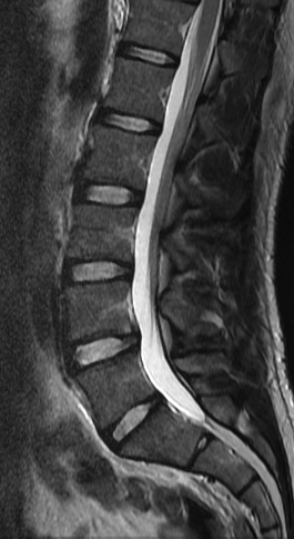
SAGITAL T2 FSE
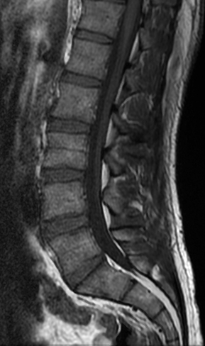
SAGITAL T1 SE
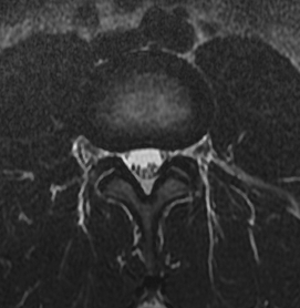
AXIAL T2 FSE
AXIALT1
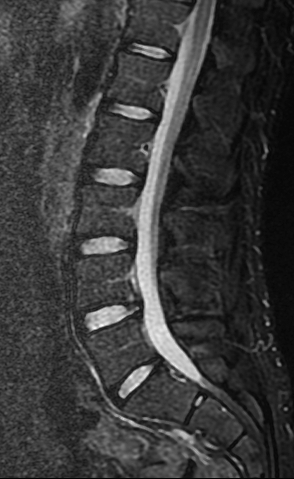
SAGITAL STIR
OPTIONAL MRI L-SPINE
CORONAL STIR
FLAIR T1 AXIAL TSE
MYELOGRAM
oblique coronal STIR through sacrum
oblique coronal T1 spin echo through sacrum
POST CONTRAST MRI L-SPINE
FAT SAT SE T1 SAGITAL
FAT SAT SE T1 AXIAL
FAT SAT SE T1 CORONAL
· Axial coverage from L3-4 through L5-S1 by default. Additional coverage more superiorly at tech’s discretion to evaluate degeneration as well.
· New optional sequences to be done only when clinician orders lumbar spine AND sacroiliac joints.
Non-contrast sagittal T1 spin echo and T2 fast spin echo, sagittal STIR, axial T1 and T2 fast spin echo through the lumbar spine. Optional oblique coronal STIR and T1 spin echo may be acquired through the sacroiliac joints.

Sagittal T2 spin echo (SE):
Particularly useful for disc spaces ve vertebral body heights and overall size of the lumbar spinal canal. Disc bulge or herniation like abnormalities, as well as he conus and filum terminale are also well visualized.
|

|
|
|
|
Normal
|
|
|
2- Sagittal T1 spin echo (SE):
Bone marrow and neural foramina are best assessed using this sequence
|

|
|
|
|
|
|
|
Normal
|
|
|
|
|
|
3- Sagittal STIR (SE)
Particularly useful to determine if there is edema in the vertebral bodies and/or disc spaces. It is a great sequence to screen the spine for subtle abnormalities.
|

|
|
|
|
Normal
|
|
|
4- Axial T2 spin echo (SE)
Useful to assess the disc and to determine if there is thecal sac or nerve root compression. The facets are also best assessed using this sequence
|

|
|
|
|
|
Normal
|
|
|
|
5- Axial T1 spin echo (SE)
Useful to evaluate the bone marrow signal and neural foramina.
AR SPINE PROTOCOL